
File:Animal Cell.svg

Size of this PNG preview of this SVG file: 800 × 462 pixels. Ither resolutions: 320 × 185 pixels | 640 × 369 pixels | 1,024 × 591 pixels | 1,280 × 739 pixels | 2,560 × 1,478 pixels | 1,405 × 811 pixels.
Oreeginal file (SVG file, nominallie 1,405 × 811 pixels, file size: 457 KB)
Ootline
| Descreeption |
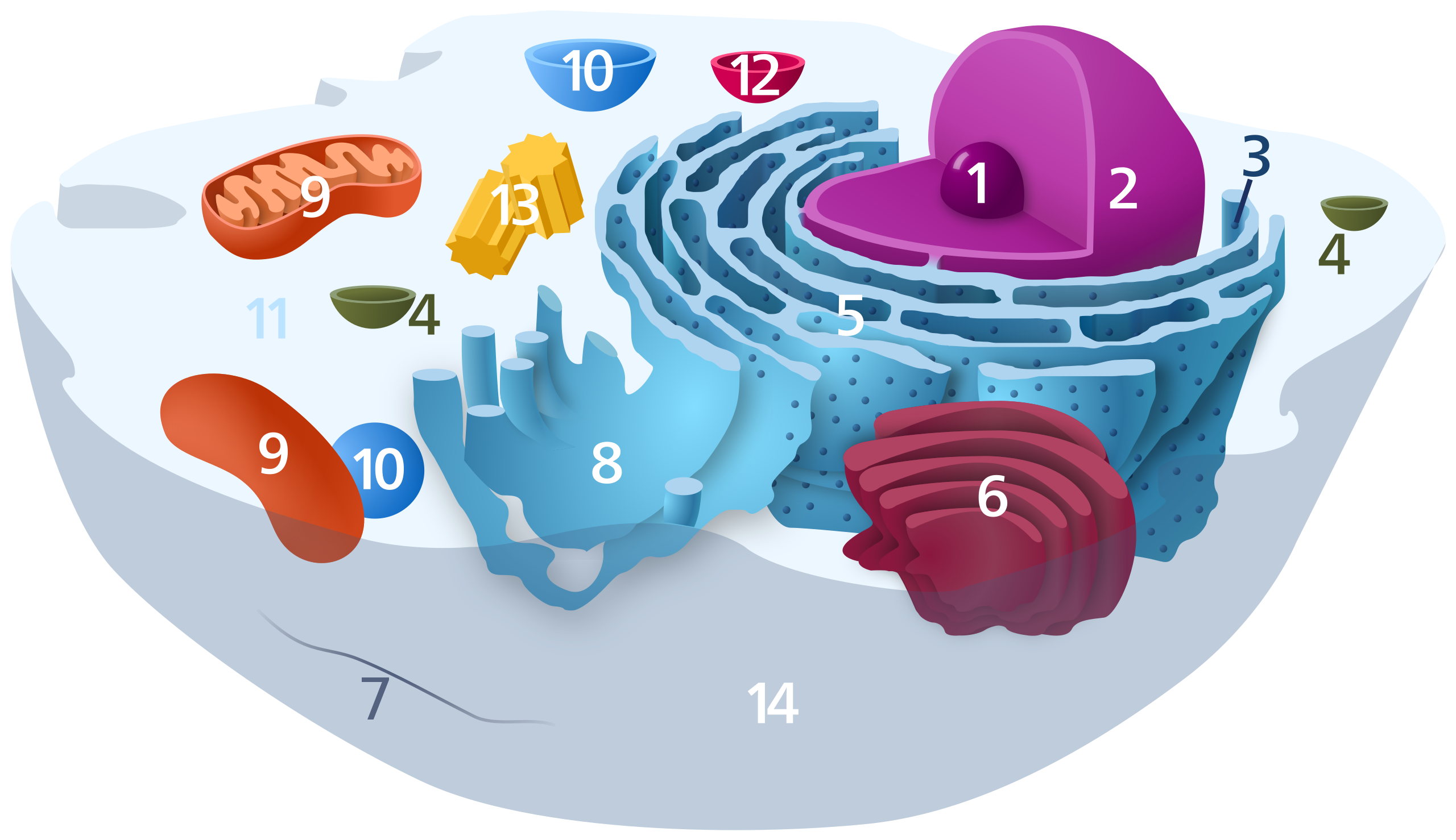
English: A reworked version of File:Biological_cell.svg.
Diagram of a typical animal cell. Organelles are labelled as follows:
العربية: رسم تخطيطي للخلية الحيوانية
Català: Dibuix esquemàtic d'una cèl·lula animal típica:
Español: Diagrama de una célula animal típica:
ਪੰਜਾਬੀ: ਕਿਸੇ ਮਿਸਾਲੀ ਜਾਨਵਰ ਦੇ ਕੋਸ਼ਾਣੂ ਦਾ ਚਿੱਤਰ:
Svenska: Schematisk bild över en typisk eukaryot cell, som visar cellens subcellulära komponenter. Organeller:
Deutsch: Organisation einer typischen eukaryotischen Tierzelle:
|
|||
| Date | ||||
| Soorce | Ain wirk | |||
| Author | Kelvinsong | |||
| Permission (Reuisin this file) |
I, the copyright holder of this work, hereby publish it under the following license:
|
File history
Clap oan ae date/time fer tae see the file aes it kithed at that time.
| Date/Time | Thummnail | Dimensions | Uiser | Comment | |
|---|---|---|---|---|---|
| current | 14:47, 17 November 2022 | | 1,405 × 811 (457 KB) | TheBartgry | Reverted to version as of 00:21, 10 December 2012 (UTC) showing continuity between nuclear membrane and ER is useful |
| 01:32, 26 Julie 2021 |  | 1,405 × 811 (452 KB) | FabPon | Reverted to version as of 00:17, 2 December 2012 (UTC) | |
| 00:21, 10 December 2012 |  | 1,405 × 811 (457 KB) | IsadoraofIbiza | Showing Nuclear membrane—ER continuity | |
| 00:17, 2 December 2012 |  | 1,405 × 811 (452 KB) | IsadoraofIbiza | center | |
| 00:07, 2 December 2012 |  | 1,466 × 891 (455 KB) | IsadoraofIbiza | Add cytoskeleton | |
| 00:03, 2 December 2012 |  | 1,466 × 891 (453 KB) | IsadoraofIbiza | User created page with UploadWizard |
Eimage airtins
The follaein pages airts tae this image:
Global file uisage
The follaein ither wikis uise this file:
- Uisage on an.wikipedia.org
- Uisage on ar.wikipedia.org
- جهاز غولجي
- ميتوكندريون
- جسيم حال
- نواة (خلية)
- ريبوسوم
- عضية خلوية
- بوابة:علم الأحياء
- هيكل خلوي
- بوابة:علم الحيوان
- بوابة:علم الأحياء/بوابات شقيقة
- شبكة إندوبلازمية
- علم الخلية
- جسم بلعمي
- نوية (خلية)
- نظام غشائي داخلي
- سيتوبلازم
- بوابة:علم الحيوان/بوابات شقيقة
- فجوة عصارية
- جسيم مركزي
- بوابة:سنوريات
- قالب:مخطط العضيات
- بوابة:سنوريات/بوابات شقيقة
- جسيم حال بلعمي
- عصارة خلوية
- قالب:مخطط العضيات/عرضي
- Uisage on bn.wikipedia.org
- Uisage on br.wikipedia.org
- Uisage on bs.wikipedia.org
- Uisage on ca.wikipedia.org
- Uisage on ckb.wikipedia.org
- Uisage on da.wikipedia.org
- Uisage on de.wikipedia.org
View mair global usage o this file.